


Led by Professor Yidong Huang, founder of Seetrum, and the research group of Associate Professor Kaiyu Cui from Tsinghua University, the team has collaborated with the Department of Ophthalmology at Peking Union Medical College Hospital and the Beijing Key Laboratory for Intelligent Diagnosis, R&D and Translation of Ophthalmic Drugs and Devices. Combining computational optics with ophthalmology, the research team has pioneered an innovative approach to spectral pathological detection for meibomian gland disorders.
Paper Title: Diagnosis of meibomian gland dysfunction based on spectral convolutional neural network chip
Authors: Yue Shi, Tianhao Liu, Shan Yang, Yu Di, Ying Li, Weihong Yu, Yidong Huang, Di Chen, Kaiyu Cui
Affiliations:
1. Department of Ophthalmology, Peking Union Medical College Hospital, Chinese Academy of Medical Sciences
2. Beijing Key Laboratory for Intelligent Diagnosis, R&D and Translation of Ophthalmic Drugs and Devices for Fundus Diseases
3. Department of Electronic Engineering, Tsinghua University
I. Research Background
Against the backdrop of intensive eye use in modern life, dry eye disease (DED) has become one of the most prevalent ocular surface disorders, affecting over 344 million people worldwide. Approximately 70% of DED cases are evaporative dry eye, primarily caused by meibomian gland dysfunction (MGD). The meibomian glands secrete meibum, which forms the outermost layer of the tear film. This lipid layer plays a vital role in suppressing tear evaporation and maintaining ocular surface homeostasis.
Current clinical diagnosis of MGD mainly relies on symptom assessment, tear film break-up time (TBUT) and other conventional examinations. These methods are highly subjective and struggle to detect early lesions. In terms of imaging technologies, infrared imaging, a widely used clinical tool, can only visualize the morphological changes of meibomian glands, yet fails to evaluate gland functions or monitor dynamic variations in meibum composition.
Spectra carry abundant information about interactions between light and matter, earning them the reputation of the "molecular fingerprint" of substances. Spectral analysis enables the identification of components within biological tissues. Traditional spectral imaging relies on point-by-point scanning across space or wavelengths, making it impossible to capture full-field spectral information in real time. Snapshot imaging technology based on metasurfaces breaks this limitation: metasurface units with distinct structures perform broadband modulation on incident light at each spatial point. After signal collection by image sensors and computational reconstruction, spectral data can be acquired, and spatial array integration realizes high-speed spectral imaging. For the first time, this study applies metasurface-based snapshot spectral imaging chips to monitor components within meibomian gland tissues, and integrates the technology with artificial intelligence (AI) algorithms to build a high-precision diagnostic framework for MGD, providing technical support for real-time, intelligent detection of ocular surface diseases.
II. Brief Introduction
This study is the first to systematically analyze the optical characteristics of tissues from MGD patients from the perspective of spectral pathology. The results reveal that MGD tissues exhibit significantly different spectral signals from healthy tissues across three key wavelength bands: 500–600 nm, 600–700 nm and 800–900 nm. These spectral variations are closely associated with abnormalities in hemoglobin derivatives and meibum components, offering new experimental evidence for understanding the mechanisms of MGD at the molecular and compositional levels.
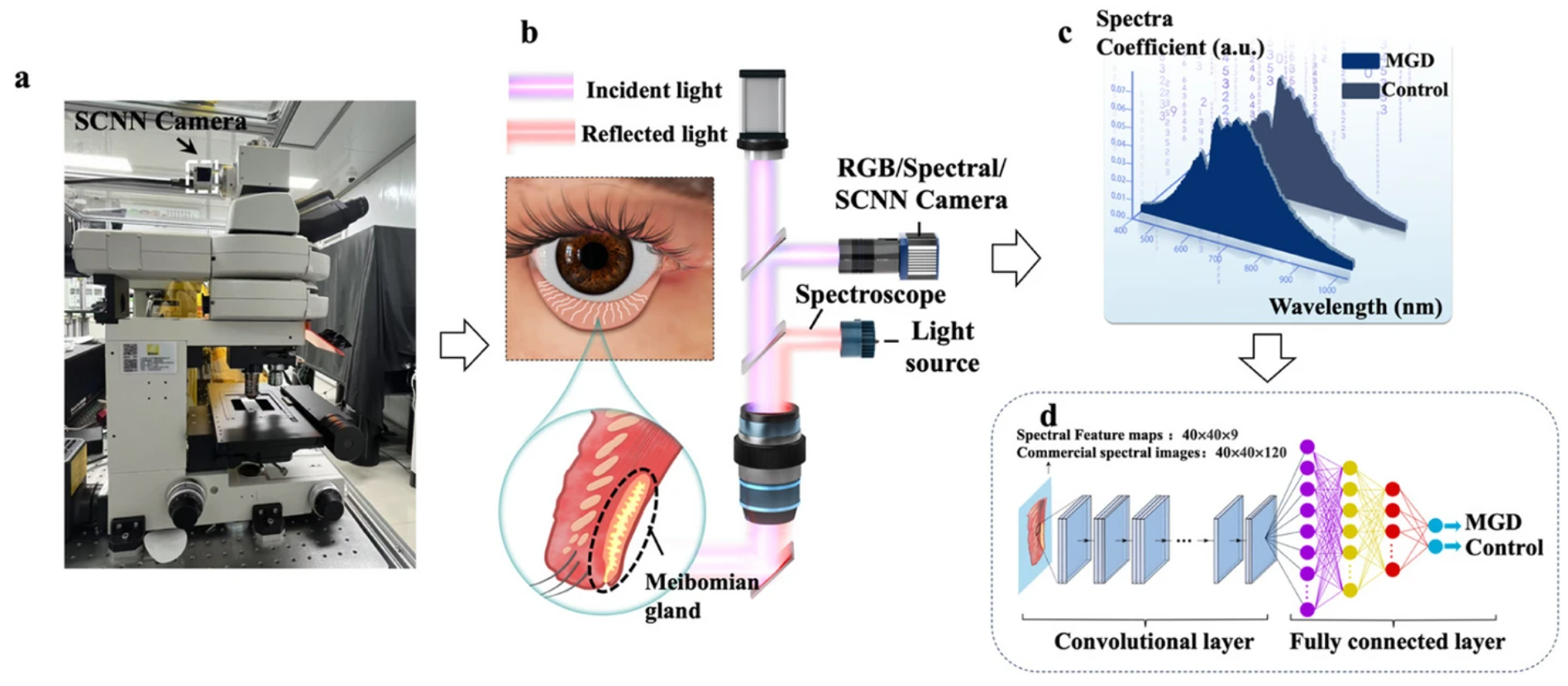
Building on this finding, the research team adopted a cutting-edge metasurface-based Spectral Convolutional Neural Network (SCNN) chip to develop an efficient intelligent diagnostic model. Deeply integrated with Complementary Metal-Oxide-Semiconductor (CMOS) image sensors, the SCNN chip eliminates mechanical scanning. It completes spectral data acquisition and on-sensor real-time computation within tens of milliseconds. Experimental results show that the SCNN model achieves a diagnostic accuracy of 96.22% for MGD, far outperforming conventional RGB image analysis with an accuracy of only 84.00%.
This research not only establishes the world’s first spectral pathological model for MGD, but also pioneers the application of optical neural network chips in the diagnosis of ocular surface diseases. It fully demonstrates the great potential of dry eye-related disorder detection to evolve toward higher precision, faster speed and miniaturization, and paves a viable technical path for non-invasive, real-time clinical screening in the future.
III. Core Advantages of the Research
This study takes metasurface spectral chips as the core technology to realize in-depth interdisciplinary integration of photonics, ophthalmology, pathological diagnosis and artificial intelligence, establishing a new all-in-one paradigm covering spectral signal acquisition and intelligent clinical interpretation.
1. Spectroscopy × Ophthalmology: From subjective assessment to objective optical characterization
Leveraging spectral imaging technology covering the 450–1000 nm wavelength range, this study identifies the unique "spectral fingerprint" of MGD-affected meibomian gland tissues for the first time. Distinct spectral differences are detected in wavelength bands corresponding to hemoglobin derivatives and meibum components. It advances the diagnosis of dry eye diseases from traditional subjective clinical evaluation and meibomian gland morphological assessment to objective, quantitative optical characterization of key tissue components.
2. Optical Neural Network × Pathological Diagnosis: On-sensor computing revolutionizes diagnostic workflows
The metasurface-based SCNN chip integrates optical convolutional layers with CMOS image sensors to adopt an on-sensor computing architecture. Without mechanical scanning, the chip synchronously completes spectral acquisition and feature extraction within tens of milliseconds, fundamentally transforming the conventional serial workflow of "data acquisition followed by post-processing computation".
3. Spectral Chip × Clinical Application: Miniaturized platforms verified for reliable diagnostic performance
Featuring compact size, low power consumption and ultra-high processing speed, the spectral chip achieves a diagnostic accuracy of 96.22% on real clinical samples. It delivers a practical technical solution for real-time, non-invasive detection and compositional analysis of meibomian gland tissues in clinical scenarios.
Summary: combining metasurface-based spectral imaging chips, on-sensor AI computing and clinical demands in ophthalmology, this research overcomes the long-standing technical bottleneck of balancing speed, accuracy and miniaturization. It represents a new direction for the paradigm shift of medical diagnosis driven by spectroscopic technologies.
IV. Main Research Contents
1. Acquisition of Spectral Feature Maps Based on the SCNN Chip
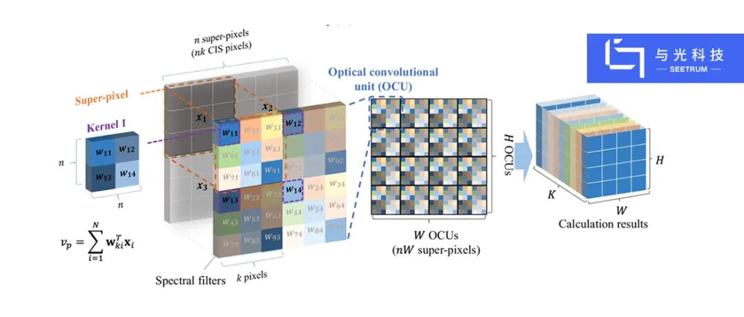
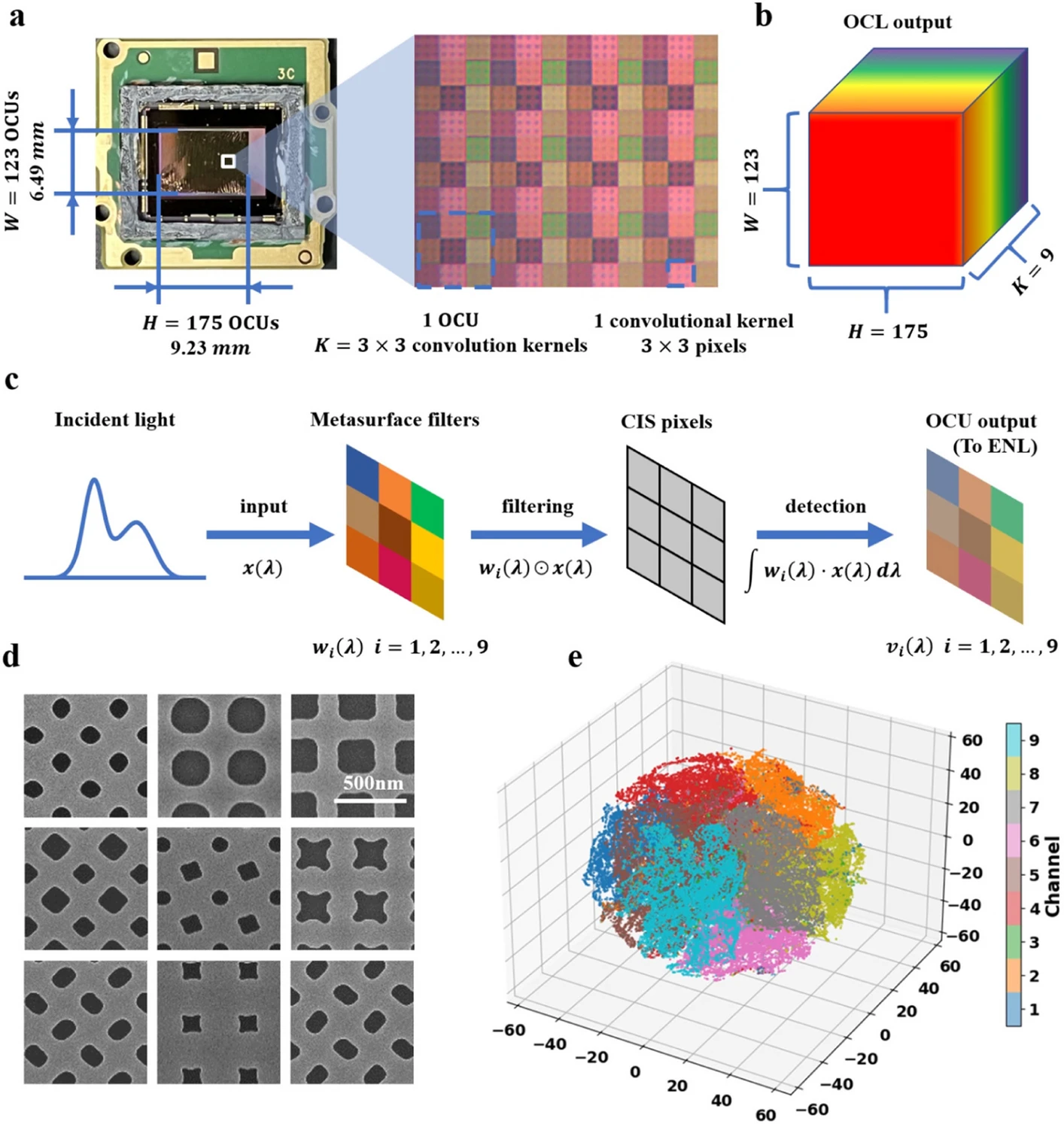
The research team used the metasurface-based SCNN chip to capture spectral feature maps of meibomian gland pathological sections under an optical microscope. The SCNN chip consists of 175 × 123 optical convolutional units, each embedded with 9 (3×3) convolution kernels, and finally outputs 3D spectral feature maps with a dimension of 175 × 123 × 9. Unlike commercial hyperspectral systems that require dozens of seconds for scanning, the SCNN chip requires no mechanical movement and finishes data collection via a single exposure, cutting the acquisition time down to tens of milliseconds.
2. Analysis of Spectral Characteristics of Meibomian Gland Tissue Components
Comparisons between the MGD group and the healthy control group reveal prominent spectral differences in key wavelength bands:
In the 600–700 nm band, the spectral coefficients of both meibomian gland regions and non-gland regions in MGD patients are significantly higher than those of the control group (P < 0.05). This band corresponds well to the absorption characteristics of hemoglobin and its derivatives (e.g., oxyhemoglobin, carboxyhemoglobin). The result indicates that MGD, a chronic inflammatory disease, may lead to changes in local tissue blood perfusion or blood oxygen saturation.
In the near-infrared band of 800–900 nm, the spectral coefficients of meibomian gland regions in MGD patients are notably lower than those of non-gland regions (P < 0.01), and significant differences also exist between the MGD group and the control group in this band (P < 0.01). This wavelength range is closely related to the light scattering properties of lipid components, which may reflect variations in light scattering caused by lipid accumulation, compositional abnormalities or structural disorders within meibomian gland acini.
3. Subgroup Analysis Verifies the Clinical Validity of Spectral Coefficients
Subgroup analysis demonstrates that groups with shorter tear film break-up time (TBUT < 10 s) present remarkably higher spectral coefficients in the 600–700 nm and 800–900 nm bands (P < 0.001). This trend is consistently validated when adopting 5 s and 8 s as cut-off values. Groups with a high Ocular Surface Disease Index (OSDI ≥ 13), poor meibomian gland expressibility (MGEX ≥ 3) and abnormal meibum quality (Meibum quality ≥ 1) also show elevated spectral coefficients in the above bands (P < 0.001).
In terms of demographic characteristics, patients aged under 50 and female patients exhibit higher spectral coefficients than their counterparts, with statistically significant differences across multiple wavelength bands. These findings align with the clinical epidemiological features of MGD, further verifying the biological relevance of spectral data.
4. Comparative Analysis of Model Performance
The spectral feature maps output by the SCNN chip were imported into electronic neural network layers for binary classification training. Meanwhile, Convolutional Neural Network (CNN) models based on commercial hyperspectral data and RGB images were established for comparison, and Monte Carlo cross-validation was adopted to evaluate overall performance.
The SCNN model achieves an average accuracy of 96.22%, an average precision of 96.34%, an average recall of 96.29% and an F1-score of 96.22%. The CNN model using commercial hyperspectral data reaches an accuracy of 95.88%, while the model based on RGB images only attains 84.00%. While maintaining diagnostic accuracy comparable to commercial hyperspectral systems, the SCNN chip boosts data acquisition speed by approximately three orders of magnitude.
V. Technological Breakthroughs and Innovations
This research delivers a series of original breakthroughs in the field of MGD diagnosis, including the establishment of a dedicated spectral database for meibomian glands, identification of characteristic spectral biomarkers, and the pioneering application of optical neural network chips in meibomian gland function diagnosis. It greatly improves diagnostic efficiency and opens up a new pathway for the precision diagnosis of ocular surface diseases.
1. World’s first spectral database for pathological sections of MGD-affected meibomian glands
This study conducts the first systematic collection and analysis of spectral data from human meibomian gland pathological tissues. The research team acquired high-quality spectral data covering the 450–1000 nm band from pathological sections of MGD patients and healthy controls, including 185 hyperspectral images, 185 SCNN spectral feature maps and 152 RGB images. Beyond recording tissue morphological information, this database captures spectral characteristics of key biochemical components such as hemoglobin and meibum, laying a solid data foundation for subsequent spectral pathological diagnostic models.
2. First identification of characteristic spectral biomarkers associated with MGD
Traditional meibomian gland imaging technologies (e.g., infrared imaging) can only observe morphological changes but cannot assess gland functions. For the first time, this study defines molecular imaging biomarkers for MGD from the spectral dimension. By comparing inherent spectral characteristics between diseased and healthy meibomian gland tissues, it identifies statistically significant and clinically reproducible spectral differences in wavelength bands corresponding to hemoglobin (reflecting inflammation and blood oxygen metabolism) and meibum (reflecting lipid composition and light scattering properties). Validated via subgroup analysis combined with multiple clinical indicators (tear film stability, symptom scores, gland secretion function, etc.), these spectral biomarkers can objectively reflect disease severity and epidemiological characteristics of different patient groups. This innovation advances MGD diagnosis from "morphological observation" to "component identification", providing brand-new biophysical targets for non-invasive, quantitative and functional assessment of ocular surface conditions.
3. Pioneering application of optical neural network chips in MGD diagnosis
Traditional hyperspectral imaging systems rely on mechanical scanning or tunable optical filters, requiring dozens of seconds for a single imaging frame. Such limitations make them unable to compensate for subtle eye movements or capture dynamic physiological processes during clinical examinations. In contrast, the metasurface-based SCNN chip is monolithically integrated with CMOS image sensors to realize on-sensor computing. Each convolution kernel on the metasurface array has a specific transmission spectrum. After performing the Hadamard product with incident light, the CIS pixels below integrate optical signals to directly output spectral feature maps. The entire process requires no scanning, and a single exposure takes only tens of milliseconds — equivalent to that of conventional RGB cameras, marking a roughly 1,000-fold increase in speed. After feature extraction by the optical convolutional layer, classification can be completed via lightweight electronic neural networks, significantly reducing the computational load on backend devices.
4. Dual breakthroughs in diagnostic accuracy and efficiency
Experimental results confirm that the SCNN model achieves a high diagnostic accuracy of 96.22%, on par with commercial hyperspectral imaging systems (95.88%), while shortening data acquisition time from dozens of seconds to tens of milliseconds. More importantly, the chip features compact size, low power consumption and compatibility with standard CMOS manufacturing processes, enabling mass production and easy integration into portable devices. This lays the groundwork for future handheld, real-time and non-invasive in-situ diagnosis of MGD, which will reshape existing clinical workflows.
VI. Conclusion and Prospects
This study systematically reveals characteristic biological markers of MGD-related tissue components from the spectral perspective, and innovatively introduces metasurface-based SCNN chips into MGD diagnosis. The SCNN chip completes spectral data collection and convolutional computation within tens of milliseconds and delivers an accuracy of 96.22%. It achieves diagnostic performance comparable to commercial hyperspectral systems with a nearly 1,000-fold increase in detection speed, and outperforms conventional RGB imaging methods by a large margin. The results fully demonstrate the unique advantages of photonic chips in medical diagnosis.
Future research will focus on the following directions: develop in-situ real-time spectral imaging technologies for ocular surfaces to translate ex-vivo pathological detection into immediate clinical testing; expand the scale of the MGD spectral database and optimize model architectures to enhance the generalization ability and stability of algorithms; extend spectral detection to the 900–1700 nm near-infrared band to further analyze the fine spectral characteristics of various lipid components in meibum; further miniaturize and system-integrate the SCNN chip to develop handheld, non-invasive and real-time diagnostic devices for MGD. Overall, this technical route is expected to drive the development of dry eye diagnosis and treatment toward higher precision, faster speed and greater intelligence, with promising prospects for clinical translation.
Relevant Links
Full paper: https://link.springer.com/article/10.1186/s43074-026-00246-2
Citation: Shi, Y., Liu, T., Yang, S. et al. Diagnosis of meibomian gland dysfunction based on spectral convolutional neural network chip. PhotoniX 7, 25 (2026). https://doi.org/10.1186/s43074-026-00246-2